![]()
Genetic of Prokaryotes and Eukaryotic Organism
Phage Phenotypes
Bacteriophage –
A bacteriophage (from ‘bacteria’ and Greek phagein, ‘to eat’) is any one of a number of viruses that infect bacteria. The term is commonly used in its shortened form, phage.
Typically, bacteriophages consist of an outer protein hull enclosing genetic material. The genetic material can be ssRNA (single stranded RNA), dsRNA, ssDNA, or dsDNA between 5 and 500 kilo base pairs long with either circular or linear arrangement. Bacteriophages are much smaller than the bacteria they destroy – usually between 20 and 200 nm in size.
T2 and its close relative T4
These are viruses that infect the bacterium E. coli. The infection ends with destruction (lysis) of the bacterial cell so these viruses are examples of bacteriophages (“bacteria eaters”).
General Phenotype –
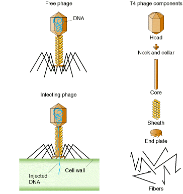
Generally each virus particle (virion) consists of:
- a protein head (~0.1 µm) inside of which is a single, circular molecule of double-stranded DNA containing 166,000 base pairs. (Figure 2.1)
- a protein tail from which extend thin protein fibers
Life Cycle –
The virus attaches to the E. coli cell. This requires a precise molecular interaction between the fibers and the cell wall of the host. The DNA molecule is injected into the cell. Within 1 minute, the viral DNA begins to be transcribed and translated into some of the viral proteins, and synthesis of host proteins is stopped. At 5 minutes, viral enzymes needed for synthesis of new viral DNA molecules are produced. At 8 minutes, some 40 different structural proteins for the viral head and tail are synthesized. At 13 minutes, assembly of new viral particles begins. At 25 minutes, the viral lysozyme destroys the bacterial cell wall and the viruses burst out — ready to infect new hosts.
- If the bacterial cells are growing in liquid culture, it turns clear.
- If the bacterial cells are growing in a “lawn” on the surface of an agar plate, then holes, called plaques (Figure 2.2), appear in the lawn.
New Phenotypes – Occasionally, new phenotypes appear such as a change in the appearance of the plaques or even a loss in the ability to infect the host.
Examples:
- h
- Some strains of E. coli, e.g. one designated B/2, gain the ability to resist infection by normal (“wild-type”) T2. The mutation has caused a change in the structure of their cell wall so that the tail fibers of T2 can no longer bind to it. However, T2 can strike back. Occasional T2 mutants appear that overcome this resistance. The mutated gene, designated h (for “host range”), encodes a change in the tail fibers so they can once again bind to the cell wall of strain B/2. The normal of “wild-type” gene is designated h+ .
- When plated on a lawn containing both E. coli B and E. coli B/2,
- the mutant (h) viruses can lyze both strains of E. coli, producing clear plaques, while
- the wild-type (h+) viruses can only lyze E. coli B producing mottled or turbid plaques.
- r
- Occasional T2 mutants appear that break out of their host cell earlier than normal.
- The mutation occurs in a gene designated r (for “rapid lysis”). It reveals itself by the extra-large plaques that it forms.
- The wild-type gene, producing a normal time of lysis, is designated r+. It forms normal-size plaques.
As with so many organisms, the occurrence of mutations provides the tools to learn about such things as
- The function of the gene;
- Its location in the DNA molecule (mapping).

Related posts:
 The Structure Analysis of Eukaryotes Introns and their Significance
 Regulation of Gene Expression in Prokaryotes and Eukaryotes
 Genetic conjugation in Bacteria
 Construction of Molecular Map
 Site Specific Recombination
 Gene Regulation in Eukaryotes
 Attenuation
 RNA Splicing