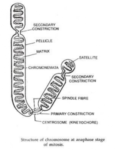
Structure of Chromosome
- For most of the life of the cell, chromosomes are too elongated and tenuous to be seen under a microscope.
- Before a cell gets ready to divide by mitosis, each chromosome is duplicated (during S phase of the cell cycle).
- As mitosis begins, the duplicated chromosomes condense into short (~ 5 µm) structures which can be stained and easily observed under the light microscope.
- These duplicated chromosomes are called dyads.

- When first seen, the duplicates are held together at their centromeres. In humans, the centromere contains ~1 million base pairs of DNA. Most of this is repetitive DNA: short sequences (e.g., 171 bp) repeated over and over in tandem arrays.
- While they are still attached, it is common to call the duplicated chromosomes sister chromatids, but this should not obscure the fact that each is a bonafide chromosome with a full complement of genes.
- The kinetochore is a complex of proteins that forms at each centromere and serves as the attachment point for the spindle fibers that will separate the sister chromatids as mitosis proceeds into anaphase.
- The shorter of the two arms extending from the centromere is called the p arm; the longer is the q arm.
Detailed structure of chromosome
- The chromosome are made up of the following parts:
- Chromatid
- Chromonema
- Centromere / Primary constriction
- NOR/ Secondary constriction –I
- Secondary Constriction II
- Telomere
- Satellite
Chromatid
At metaphase Chromosome are made up of two thread like structure which is called chromatid. These chromatids are joined at a place which is called centromere. These are separated in Anaphase stage. They carry by spindle fiber on the opposite pole .These are two type
1- Sister chromatid
At the start of meiosis each chromosome is replicate (s-stage) and make two identical copies of DNA which are attached to each other at centromere. These are called sister chromatid.
2-Non sister chromatid
Two chromatid of two homologous chromosome are known as non sister chromatid.
In these one chromatid come from father and one chromatid come form mother in the progeny.
Chromonema –
During prophase and also interphase, the chromosomal material becomes visible as a very thin filament which are called chromonemata and which represents chromatid in early stages of condensation. It is a single linear DNA molecule with its associated proteins.
Centromere/Primary constriction –
- Each chromosome have a groove which is known as centromere.
- In this region Kinetochore protein are present. In which spindle fiber are attached which carry chromosome to opposite pole.
- The centromere lies within a thinner segment of the chromosome, the primary constriction.
- The regions flanking the centromere frequently contain highly repetitive DNA and stain more intensely with basic dyes (heterochromatin).
- Centromere contains specific DNA sequences with special protein bound to them forming a disc shaped structure called as kinetochore.
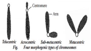
According to the no. of centromere these are the following types
- 1- Monocentric – Only single centromere present
- 2- Di-centric – Two centromere present
- 3- Poly centric – More than two centromere are present.
- 4- Acentric – Centromere absent
According to the position of centromere chromosomes are following types
1. Telocentric –
- In this centromere are present on the extreme end (head)
- This chromosome contain only one long arm.
- These are rod shaped and are present only in plants
2. Acrocentric
- In this type of chromosome centromere is positioned sub terminally.
- So this chromosome have a very small arm and other large arm
3. Sub metacentric
- In this type of chromosome centromere is present sub median position of chromosome
- So it have unequal arm
- it is look like “J” , or “L” shape.
4. Metacentric
- In this type of chromosome the centromere is present at middle point of chromosome
- So it have two equal arm.
- It is have “V” like shape
4. Kinetochore –
5. Chromomere –
These components are beads like accumulation of chromatin material that are sometimes visible along interphase chromosomes. Chromomeres are especially present in polytene chromosomes where they become aligned side by side, constituting the chromosome bands. These tightly folded regions of DNA are of great interest, as they may correspond to the units of genetic functions in the chromosome. At metaphase, chromomeres are not visible as chromosome is tightly coiled.
6. Telomere –
This term refers to the tips of the chromosomes. Telomeres have special properties that when chromosomes are broken, the free end without telomeres fuse with other broken chromosomes; however they do not fuse with a normal telomere. Telomeres contain the ends of the long linear DNA molecule contain in each chromatid and therefore have an unusual DNA structure.
- The end part of the chromosome called telomere.
- In this region chromatin is present in the form of Loop which make heterochromatic part.
- It avoid to join of chromosome.
- It is formed by the help of telomerase enzyme.
- In this aprx. 10kb repetitive sequence are present.
- In this one end have “G” and another end have “C” as residue on it.
- Protein that Bind to the repetitive sequence of telomeric DNA is Ta2/P protein.
- Two type of protein Ku & Sir are help in replication of Telomeric DNA and also control the Length of this Part
7. Nucleolar Organizers –
A secondary constriction in the chromosome is a constriction other than the centromere. It is often associated with the nucleolus during interphase and may take part in the reorganization of the nucleolus at the end of cell division. Secondary constrictions are constant in their position and extents, and therefore useful in identifying particular chromosomes in a set. Secondary constrictions are distinguished from the primary constriction by the absence of marked angular deviation of the chromosomal segments during anaphase.
- Another constriction of the chromosome is called secondary constriction or NOR
- It’s role in forming of Nucleolus
- A round structure is present on it which is called satellite
- In human it is present on the Chs no. 13,14,15,21 &22
These areas are certain secondary constriction that contains the gene coding for 18S and 28S ribosomal RNA and that induce the formation of nucleoli.
8. Secondary constrictions II
- Some time other than secondary constriction I another constriction is present on the chromosome which is called secondary constriction II (SCII)
- It is present on the specific place which help in recognization of chromosome .
- In human it is present on the Chs No. 1,10,13,16 &Y
9. Satellites –
- A rounded button like structure is present ahead to the secondary constriction which is known as satellite.
- It attached to the chromosome by a thin thread like structure
- The chromosome on which it attaches is called SAT chromosme
- The satellite and constriction are constant in shape and size for each particular chromosome.