![]()
PACKAGING OF DNA
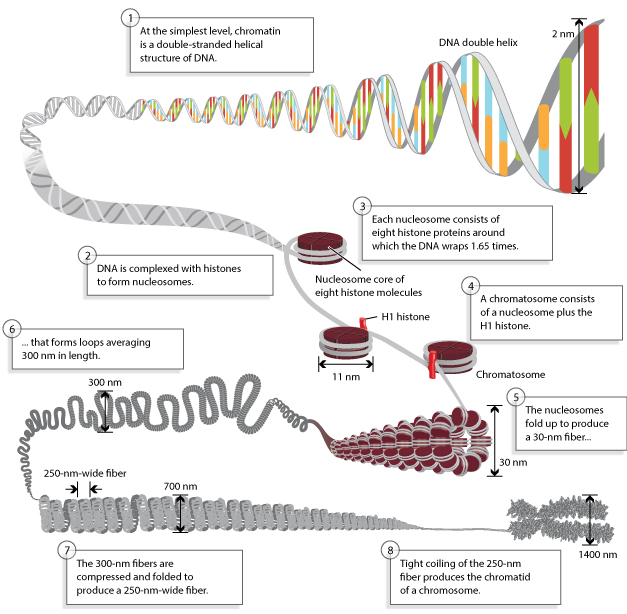
- In living organisms, the length of DNA always greatly exceeds the dimensions of the nucleus which contains it.
- The DNA must be compressed very tightly to fit into the available space.
- When comparing prokaryotic cells to eukaryotic cells, prokaryotes are much simpler than eukaryotes in many of their features.
- Most prokaryotes contain a single, circular chromosome that is found in an area of the cytoplasm called the nucleoid.
- The size of the genome in one of the most well-studied prokaryotes, E.coli, is 4.6 million base pairs (approximately 1.1 mm, if cut and stretched out).
- So how does this fit inside a small bacterial cell?
- The DNA is twisted by what is known as supercoiling. Supercoiling means that DNA is either under-wound (less than one turn of the helix per 10 base pairs) or over-wound (more than 1 turn per 10 base pairs) from its normal relaxed state.
- Some proteins are known to be involved in the supercoiling; other proteins and enzymes such as DNA gyrase help in maintaining the supercoiled structure.
- The overall compression of the DNA has also been described as packaging ratio, means the length of DNA divided by the length of unit that contain it.
- Eukaryotes, whose chromosomes each consist of a linear DNA molecule, employ a different type of packing strategy to fit their DNA inside the nucleus.
- The ratio of salivary gland chromosome of Drosophila at metaphase is 100:1.Each chromatid, therefore, consists of a single unbroken molecule of DNA which is coiled, supercoiled and folded to form the chromatid.
- Compaction is aided by the associated nucleoprotein
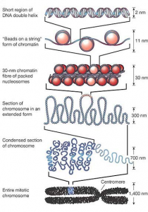
Nucleosome
- When Chromosomes are lysed gently, a 250 Ǻ thick chromatin network is obtained.
- After treatment with reagents like EDTA or sodium citrate, which remove the calcium ions, 100 Ǻ thick fibres are obtained, indicating thereby that each 250 Ǻ thick fibres is composed of two 100 Ǻ.
- The 100 Ǻ fibres are composed of DNA and protein. When the protein portion is digested by pronase, the DNA double helix is exposed.
- In conjunction with histones the DNA duplex contracts a great deal and becomes considerably thick to form the 100 Ǻ fibre.
- The 250 Ǻ fibres, which are sometimes branched, are perhaps formed due to the foldings of a 100 Ǻ fibre.
- How the 250 Ǻ fibers are organized to form the chromosomes is still a mystery, but the organization of 100 Ǻ fibers is more or less known.
- Olius and Olius (1973) and woodcock (1973) exposed isolated nuclei to hypotonic buffers and centrifuged them through a formaldehyde solution onto a grid.
- When seen through the electron microscope, such preparations appeared as beads on a string.
- They suggested that the beaded structure represents highly compact particles of DNA and histones in a regular repeating array.
- These particles, 70 to 90 Ǻ across, were later called nucleosome by Oudet et al. (1975).
- Nucleosome (or nu bodies) have now been crystallized and subjected to neutron-scattering, X-ray diffraction, nuclease digestion and high resolution electron microscopy.
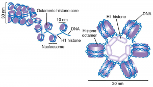
- These studies suggest that four out of the five types of histone molecules found in eukaryotic cells, associate with each other to form dimers, which in turn associate to form an octamer.
- The DNA double helix, about 140 nucleotide pairs long, is wrapped around the octamer, forming approximately 1¾ turns around the histone octamer.
- This core particle is discoid or oblate and adjacent particles are connected by the intercore DNA helix.
- Treatment of chromatin with micrococcal nuclease cuts the connecting double-stranded DNA to generate nucleosomal monomers, each containing about 160 base pairs of DNA.
- Further digestion removes the spacer helix and results into the core nucleosome with about 140 base pairs.
- The size of DNA fragments from diverse cell types indicates that the nucleosome structure is the same whereas there is a wide variation in the size of the spacer.
- Prolonged digestion results into subnucleosomal particles.
Chromatin → Nucleosomal monomers Containing the spacer DNA (160 base pairs) → Core nucleosomes (140 base pairs) → Subnucleosomal particles
- Four (H2A, H2B, H3, and H4) out of the five types of histones are associated with the nucleosome core.
- Equimolar amounts of these four inner histones can be used to reconstruct nucleosomes.
- Mixtures of H3 and H4 also permit the organization of nucleosome-like particles, but individual histones or mixtures of H2A and H2B fail to do so. Thus, H3 and H4 appear to play a crucial role in the organization of nucleosomes.
- The 140 base pair long DNA associated with the core nucleosome is frequently cut by DNase I at approximately 20, 40, 50, 100, 120 and 130 bases from the 5’ end, indicating thereby that these are the sites which are not in close association with the inner histones and are therefore more prone to nuclease digestion.
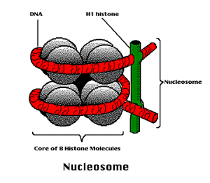
- H1 has been found to be associated with the spacer region.
- The size of H1 histone and its content of basic amino acids control the length of the spacer DNA.
- It is believed to control the super-nucleosomal organization (e.g. coiling and supercoiling of the helix) in the chromosomes.
- The 200-300 Ǻ wide chromatin fibres become thinner after the removal of H1 histones and reagents which bring about cross-linking result into polymerization of H1 molecules.
- It has been suggested that H1-H1 contacts are possibly responsible for the stabilization of the 200-300 Ǻ thick chromatin thread.
- As soon as these contacts are severed by the removal or modification of H1 histones, the nucleosomal beads on a string can be clearly seen.
- Nucleosome has a packaging function, organizing the constituent DNA into a structure about six fold shorter than the DNA duplex.
- Folding of these DNA results into a close-packed alignment of nucleosomes and shortens the DNA by another factor of 5 or 10 into 200-300 Ǻ thick fibres.
- The entire eukaryotic DNA, whether transcriptionally active or inactive, is organized into nucleosomes.
- Chromatin structure and nucleosomal conformation is altered in some unknown way at the time of transcription.
- This is essential to expose the DNA for RNA synthesis.