![]()
Thickening of cell wall
- When process of cell wall formation Is completed and cell obtains iIts full shape and size thereafter lignification of cell wall starts
- This lignification process does not occur in every cell but happens In those cells which are either Involved In transport or provide mechanical strength for example. vessels. tracheid and sclerenchyma etc.
- Deposition of cell wall is possible only by lignin. This process is called lignification.
- Lignin accumulated in sclerenchymatous cells and form a complete lignified laver
- On the basis of type of Lignification. the following different types of structures are found In vessels and tracheids
(A). Annular :
- In this Lignin is deposited in the form of a ring
- It is often present In protoxylem of tracheids and vessels.
(B) Spiral :
- In this type of thickening of lignin is present in the form of a spiral band in protoxylem.
(C) Scalariform :
- In this type of thickening lignin Is deposited as transverse bands of the ladder on Inner surface of the primary cell wall.
- Such lignified bands or ‘rods are present parallel to each other and at right angle to the long axis of the cell.
- It looks Like a scalariform ( ladder like)
(D) Reticulate :
- In this lignin bands are organised in the form of interconnected that form a net like structure which is formed on the Inner surface of cell wall which ‘said to be reticulate thickening.

(E) Pitted :
- In this Lignin is ‘deposited evenly on wall and the entire wall is lignified leaving some small ‘spaces such lignin free spaces are called pits.
- This type of lignification is called pitted thickening.
These pits may be of two types
Simple pits :
- These simple pits are seen as round/oval when observed under the microscope.
- In this the whole area of simple pit can be seen up to its complete depth
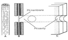
- These pits are found at the same place on the adjacent walls of two cells because these pts are present In pairs.
- Only the middle lamella separates these pits pairs. Hence this middle lamella is also called as pit membrane or closing membrane.
- Simple pits are seen in living cells such as parenchyma and collenchyma. Protoplasm of two adjacent cells are exchanged through these Simple pits.
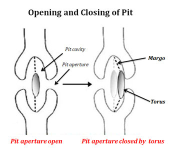
Bordered pits :
- In bordered pit the diameter of depth between cell wall and pit membrane Increases inside.
- In the bordered pit secondary wall is present therefore each pit appears as a funnel shaped structure in each pit pair of two adjacent walls the broad part of both the funnels are kept facing each other. The pit membrane is present in between them.
- The Pits are actually the site for diffusion between two cells from which liquid material can be exchanged
- In this type of pits a closing membrane or pit membrane is also thickened it is called torus.
- When the diffusion stops, the torous pressed toward the pit and closes the passage, so it controls the diffusion process.
- Bordered pits are found mainly in tracheids and vessels of gymnosperms. They are also found in major quantities in fibres, stone cells. wood parenchyma, companion cells and sieve tubes.