![]()
Lysosomes
- The lysosomes (Gr. lyso=digestive + soma = bodies) are tiny membrane-bound vesicles involved in intracellular digestion.
- They contain a variety of hydrolytic enzymes that remain active under acidic conditions.
- The lysosomal lumen is maintained at an acidic pH (around 5) by an ATP-driven proton pump in the membrane.
- Thus, these remarkable organelles are primarily meant for the digestion of a variety of biological materials and secondarily cause aging and death of animal cells and also a variety of human diseases such as cancer, gout Pompe’s silicosis and I-cell disease.

History
- During early electron microscopic studies, rounded dense bodies were observed in rat liver cells.
- These bodies were initially described as “perinulclear dense bodies”,
- C. de Duve, in 1955, renamed these organelles as “lysosomes” to indicate that the internal digestive enzymes only became apparent when the membrane of these organelles was lysed.
- However, the term lysosome means lytic body having digestive enzymes capable of lysis (viz., dissolution of a cell or tissue.
Lysosomes were investigated according to following two schools
- (1) C, de Duve and his coworkers (1963, 1964) worked in Belgium and their approach was biochemical one .
- (2) Alex Novikoff and his research group (1962, 1964) worked in United States and their approach was morphological and cytochemical.
- For the discovery of lysosomes and a brilliant series of experiments on them, de Duve shared the 1974 Nobel Prize for physicality with Palade andClaude, both were pioneer cells biologists.
Occurrence
- The lysosomes occur in most animal and few plant cells.
- They are absent in bacteria and mature mammalian erythrocytes.
- Few lysosomes occur in muscle cells or in cells of the pancreas.
- Leucocytes, especially granulocytes are a particularly rich source of lysosomes.
- Their lysosomes are so large that they can be observed under the light microscope.
- Lysosomes are also numerous in epithelial cells of absorptive, secretory and excretory organs (e.g., intestine, liver, kidney, etc.)
- They occur in abundance in the epithelial cells of lungs and uterus.
- Lastly phagocytic cells and cells of the reticuloendothelial system (e.g., bone marrow, spleen and liver) are also rich in lysosomes.
Structure
- The lysosomes are round vacuolar structures which remain filled with dense material and are bounded by a single unit membrane.
- Their shape and density vary greatly.
- Lysosomes are 0.2 to 0.5mm in size.
- Since, size and shape of lysosomes vary from cell to cell and from time to time (i.e. they are polymorphic), their identification becomes difficult.
- However, on the basis of the following three criteria, a cellular entity can be identified as a lysosome :
- It should be bound by a limiting membrane
- It should contain two or more acid hydrolases ; and
- It should demonstrate the property of enzyme latency when treated in a way that adversely affects organelle’s membrane structure.
Isolation and Chemical Composition
- Lysosomes are very delicate and fragile organelles.
- Lysosomal fractions have been isolated by sucrose-density centrifugation (or Isopycnic centrifugation) after mild methods of homogenization.
- Since the original de Duve’s isolated lysosomal fractions were having contaminations of mitochondria, microsomes and microbodies, so, 1960’s it was investigated that rats injected with dextran or Triton WR-1339, incorporated these compounds into their lysosomes, thereby altering their density and making their cleaner separation possible by differential centrifugation and density gradients (see Reid and Leech, 1980).
- Lysosomes tend to accumulate certain dyes (vital stains such as Neutral red, Niagara, Evans blue) and drugs such as anti-malarial drug chloroquine.
- Such ‘loaded’ lysosomes can be demonstrated by fluorescence microscopy.
- The location of the lysosomes in the cell can also be pinpointed by various histochemical or cytochemical methods.
- For example, lysosomes demonstrate the property of metachromasia with toluidine blue and give a positive acid Schiff reaction
- Metachromasia is the property exhibited by certain pure dyestuffs, chiefly basic stains, of coloring certain tissue elements in a different colour.
- Certain lysosomal enzymes are good histochemical markers.
- For example, acid phosphatase is the principal enzyme which is used as a marker for the lysosomes by the use of Gomori staining technique.
- Specific stains are also used for other lysosomal enzymes such as B-glucuronidase, arylsulphatase, N-acetyl-B-glucosaminidase and 5-bromo-4-chloro indole acetate esterase.
Lysosomal Enzymes
- According to a recent estimate, a lysosome may contain up to 40 types of hydrolytic enzymes. they include proteases (e.g Cathepsin for protein digestion), nucleases, glycosidases (for digestion of polysaccharides and glycosides ), lipase, phospholipases, phosphatases and sulphatases.
- All lysosomal enzymes are acid hydrolases, optimally active at the pH5 maintained within lysosomes.
- The lysosomal enzymes latent and out of the cytoplasmic matrix or cytosol (whose pH is about ~ 7.2), but the acid dependency of lysosomal enzymes protects the contents of the cytosol (cytoplasmic matrix) against any damage even if leakage of lysosomal enzymes should occur.
- The so-called latency of the lysosomal enzymes is due to the presence of the membrane which is resistant to the enzymes that it encloses.
- Most probably this is due to the fact that most lysosomal hydrolases are membrane-bound, which may be prevalent in the active center of enzymes to gain access to susceptible groups in the membrane.
Kind of Lysosomes
- Lysosomes are extremely dynamic organelles, exhibiting polymorphism in their morphology.
- Following four types of lysosomes have been recognized in different types of cells or at different time in the same cell.
- Of these, only the first is the primary lysosome, the other three have been grouped together as secondary lysosomes.

1. Primary Lysosomes
- These are also called storage granules, proto lysosomes or virgin lysosomes.
- Primary lysosomes are newly formed organelles bounded by a single membrane and typically having a diameter of 100 nm.
- They contain the degradative enzymes which have not participated in any digestive process.
- Each primary lysosome contains one type of enzyme or another and it is only in the secondary lysosome that the full complement of acid hydrolases is present.
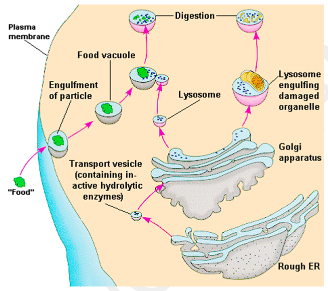
2. Heterophagosome
- They are also called heterophasic vacuoles, hetero lysosomes or phagolysosomes.
- Hetero Phagosomes are formed by the fusion of primary lysosomes with cytoplasmic vacuoles containing extracellular particles into the cell by any of a variety of endocytic processes (e.g., pinocytosis, phagocytosis or receptor mediated endocytosis.
- The digestion of engulfed substances takes place by the enzymatic activities of the hydrolytic enzymes of the secondary lysosomes.
- The digested material has low molecular weight and readily passes through the membrane of the lysosomes to become the part of the matrix.
3. Autophagosomes
- They are also called autophagic vacuole, cyto lysosomes or autolysosomes.
- Primary lysosomes are able to digest intracellular structures including mitochondria, ribosomes, peroxisomes and glycogen granules.
- Such autodigestion (called autophagy) of cellular organelles is a normal event during cell growth and repair and is especially prevalent in differentiating and de-differentiating tissues (e.g., cells undergoing programmed death during meta-morphosis or regeneration) and tissue under stress.
- Autophagy takes several forms.
- In some cases the lysosome appears to flow around the cell structure and fuse, enclosing it in a double membrane sac, the lysosomal enzymes being initially confined between the membrane.
- The inner membrane then breaks down and the enzymes are able to penetrate to the enclosed organelle.
- In other cases, the organelle to be digested is first encased by smooth ER, forming a vesicle that fuses with a primary lysosome.
- Lysosomes also regularly engulf bits of cytosol (cytoplasmic matrix) which is degraded by a process called microautophagy.
- As digestion proceeds, it becomes increasingly difficult to identify the nature of the original secondary lysosome (i.e., heterophagosome or autophagosome) and the more general term digestive vacuole is used to describe the organelle at this stage.
4. Residual Bodies
- They are also called telo lysosomes or dense bodies.
- Residual bodies are forced if the digestion inside the food vacuole is incomplete. Incomplete digestion may be due to absence of some lysosomal enzymes.
- The undigested food is present in the digestive vacuole as the residues and may take the form of whorls of membranes. grains, amorphous masses, ferritin-like or myelin figures.
- Residual bodies are large, irregular in shape and are usually quite electron-dense. In some cells, such as Amoeba and other protozoa, these residual bodies are eliminated.
- In other cells, residual bodies may remain for a long time and may load the cells to result in their aging.
- For example, pigment inclusions (age pigment or lipofuscin granules) found in nerve cells (also in liver cells, heart cells and muscle cells) of old animals may be due to the accumulation of residual bodies.
Origin
- The biogenesis (origin) of the lysosomes requires the synthesis of specialized lysosomal hydrolases and membrane proteins.
- Both classes of proteins are synthesized in the ER and transported through the Golgi apparatus, then transported from the trans Golgi network to an intermediate compartment (an endolysosome) by means of transport vesicles (which are coated by clathrin protein).
- The lysosomal enzymes are glycol proteins, containing N-linked oligosaccharides that are processed in a unique way in the cis Golgi so that their mannose residues are phosphorylated.
- These mannose 6-phosphate (M6P) groups are recognized by M6P groups are recognized by M6P-receptors (which are transmembrane proteins) in the trans Golgi network that segregates the hydrolases and helps to package them into budding clathrin-coated vesicles which quickly lose their coats.
- These transport vesicles containing the M6P-receptors act as shuttles that move the receptors back and forth between the Golgi network and endolysosomes.
- They low pH in the endolysosome dissociates the lysosomal hydrolases from this receptor, making the transport of the hydrolases unidirectional.
Function of Lysosomes
The important functions of lysosomes are as follow :
1. Digestion of large extracellular particles.
- The lysosomes digests the food contents of the phagosomes or pinosomes.
2. Digestion of intracellular substances.
- During starvation, the lysosomes digest the stored food contents viz. proteins, lipids and carbohydrates (glycogen) of the cytoplasm and supply to the cell necessary amounts of energy.
3. Autolysis.
- In certain pathological conditions the lysosomes start to digest the various organelles of the cells and this process is known as autolysis or cellular autophagy.
- When a cell dies, the lysosome membrane ruptures and enzymes are liberated.
- These enzymes digest the dead cells.
- In the process of metamorphosis of amphibians and / tunicates many embryonic tissues, e.g., gills, fins, tail, etc., are digested by the lysosomes and utilized by the other cells.
4. Extracellular Digestion.
- The lysosomes of certain cells such as sperms discharge their enzymes outside the cell during the process of fertilization.
- The lysosomal enzymes digest the limiting membrane of the ovum and form a penetration path in the ovum for the sperms.
- Acid hydrolases are released from osteoclasts and break down bone for the reabsorption ; these cells also secrete lactic acid which makes the local pH enough for optimal enzyme activity.
- Likewise, preceding ossification (bone formation), fibroblasts release cathepsin D enzyme to break down the connective tissue.
Lysosomes in plants
- Plants contain several hydrolases, but they are not always as neatly compartmentalized as they are in animal cells.
- Many of these hydrolases are found bound to and functioning within the vicinity of the cell wall and are not necessarily contained in membrane bound vacuoles at these sites.
- Many types of vacuoles and storage granules of plants are found to contain certain digestive enzymes and these granules are considered as lysosomes of plant cell.
- According to Matile the plant lysosomes can be defined as membrane bound cell compartments containing hydrolytic digestive enzymes.
- Matile has divided vacuoles of plants into following three types :
1. Vacuoles
- The vacuole of a mature plant cell is formed from the enlargement and fusion of smaller vacuoles present in meristematic cells; these pro vacuoles, which are believed to be derived from the ER and possibly the Golgi and contain acid hydrolases.
- These lysosomal enzymes are associated with the tonoplast of large vacuoles of differentiating cells.
- Sometimes, mitochondria and plastids are observed inside the vacuole suggesting autophagy in plants.
2. Spherosomes
- The spherosomes are membrane bounded, spherical particles of 0.5 to 2.5 mm diameter, occurring in most plant cells.
- They have a fine granular structure internally which is rich in lipids and proteins.
- They originate from the endoplasmic reticulum (ER).
- Oil accumulates at the end of a strand of ER and a small vesicle is then cut off by contribution to form particles, called prospherosomes.
- The prospherosomes grow in size to form spherosomes.
- Basically, the spherosomes are involved in lipid synthesis and storage. But, the spherosomes of maize root tips and spherosome of tobacco endosperm tissue (Spichiger, 1969) have been found rich in hydrolytic digestive enzymes and so have been considered as lysosomes.
- Like lysosomes they are not only responsible for the accumulation and mobilization of reserve lipids, but also for the digestion of other cytoplasmic components incorporated by phagocytosis.
3. Aleurone Grain
- The aleurone grains or protein bodies are spherical membrane-bounded storage particles occurring in the cells of endosperm and cotyledons of seeds.
- They are formed during the later stages of seed ripening and disappear in the early stages of germination.
- They store protein (e.g., globulins) and phosphate in the form of phytin.
- Matile has demonstrated that aleurone grains from pea seed contain a wide range of hydrolytic enzymes including protease and phosphatase which are required for the mobilization of stored protein and phosphate, although the presence of other enzymes such as b-amylase and RNAse suggest that other cell constituents may also be digested.
- Thus like spherosomes, aleurone grains store reserve materials, mobilize them during germination and in addition form a compartment for the digestion of other cell components.
- The aleurone grains are derived from the strands of the endoplasmic reticulum.
- During germination of barley seed, the activity of hydrolases is found to be controlled by hormones such as gibberellic acid.
- Gibberellic acid, a plant growth hormone, is released by the embryo to the aleurone layer where, in turn, the hydrolases are released to the endosperm.
- This hormone operates by derepressing appropriate genes in the aleurone cells, which then begin to crank out new hydrolytic proteins .