![]()
Golgi Apparatus
- For the performance of certain important cellular functions such as biosynthesis of polysaccharides, packaging (compartmentalizing) of cellular synthetic products (proteins), production of exocytotic (secretory) vesicles and differentiation of cellular membranes, there occurs a complex organelle called Golgi complex or Golgi apparatus in the in the cytoplasm of animal and plant cells.
- The Golgi apparatus, like the endoplasmic reticulum, is a canalicular system with sacs, but unlike the endoplasmic reticulum it has parallely arranged, flattened, membrane-bounded vesicles which lack ribosomes and stainable by osmium tetraoxide and silver salts.

History
- An Italian neurologist (i.e., physician) Camillo Golgi in 1873 discovered and developed the silver chromate method (termed la reazione nera) for studying histological details of nerve cells. of cerebral cortex of brain) of barn owl contained and internal reticular network which stains black with the silver stain.
- He called this structure apparato reticolare interno (internal reticular apparatus).
- By reporting the existence of such an organelle inside cell, he inadvertently raised a storm of controversy in the scientific world, which is commonly known as the Golgi controversy.
Occurence
- The Golgi apparatus occurs in all cells except the prokaryotic cells (viz., mycoplasmas, bacteria and blue green algae) and eukaryotic cells of certain fungi, sperm cells of bryophytes and pteridiophytes, cells of mature sieve tubes of plants and mature sperm and red blood cells of animals.
- Their number per plant cell can vary from several hundred as in tissues of corn root and algal rhizoids (i.e., more than 25,000 in algal rhizoids,), to a single organelle in some algae.
- Certain algal cells such as Pinularia and Microsterias, contain largest and most complicated Golgi apparatuses.
- In higher plants, Golgi apparatuses are particularly common in secretory cells and in young rapidly growing cells.
- In animal cells, there usually occurs a single Golgi apparatus, however, its number may vary from animal to animal and from cell to cell.
- Thus Paramoeba species has two Golgi apparatus and nerve cells, liver cells and chordate oocytes have multiple Golgi apparatuses, there being about 50 of them in the liver cells.
Distribution
- In the cells of higher plants, the Golgi bodies or dictyosomes are usually found scattered throughout the cytoplasm and their distribution does not seem to be ordered or localized in any particular manner
- However, in animal cells the Golgi apparatus is a localized organelle.
- For example, in the cells of ectodermal or endodermal origin, the Golgi apparatus remains polar and occurs in between the nucleus and the periphery (e.g., thyroid cells, exocrine pancreatic cells and mucus-producing goblet cells of intestinal epithelium) and in the nerve cells it occupies a circum-nuclear position.
Morphology
- The Golgi apparatus is morphologically very similar in both plant and animal cells.
- However, it is extremely pleomorphic: in some cell types it appears compact and limited, in others spread out and reticular (net-like).
- Its shape and form may very depending on cell type.
- Typically, however, Golgi apparatus appears as a complex array of interconnecting tubules, vesicles and cisternae.
- There has been much debate concerning the terminology of the Golgi’s parts.
- The classification given by D.J. Morre (1977) is most widely used.
- In this scheme, the simplest unit of the Golgi apparatus is the cisterna.
- This is a membrane bound space in which various materials and secretions may accumulate.
- Numerous cisternae are associated with each other and appear in a stack like (lamellar) aggregation.
- A group these cisternae is called the dictyosome, and a group of dictyosomes makes up the cells Golgi apparatus.
- All dictyosomes of a cell have a common function
1.Flattened Sac or Cisternae
- Cisternae (aboute 1mm in diameter) are central, flattened, plate-like or saucer-like closed compartments which are help in parallel bundles or stacks on above the other.
- In each stack, cisternae, are separated by a space of 20 to 30 nm which may contain rod-like elements or fibres.
- Each stack of cisternae forms a dictyosome which may contain 5 to 6 Golgi cisternae in animal cells or 20 or more cisternae in plant cells.
- Each cisterna is bounded by a smooth unit membrane (7.5 nm thick), having a lumen varying in width from about 500 to 1000 nm.
- The margins of each cisterna are gently curved so that the entire dictyosome of Golgi apparatus takes on a bow like appearance.
- The cisternae at the convex end of the dictyosome comprise proximal forming or cis-face and the cisternae at the concave end of the dictyosome comprise the distal, maturing or trans-face.
- The forming or cis-face of Golgi is located next to either the nucleus or a specialized portion of rough ER that lacks bound ribosomes and is called ”transitional” ER.
- Trans face of Golgi is located near the plasma membrane.
- This polarization is called cis-trans axis of the Golgi apparatus.
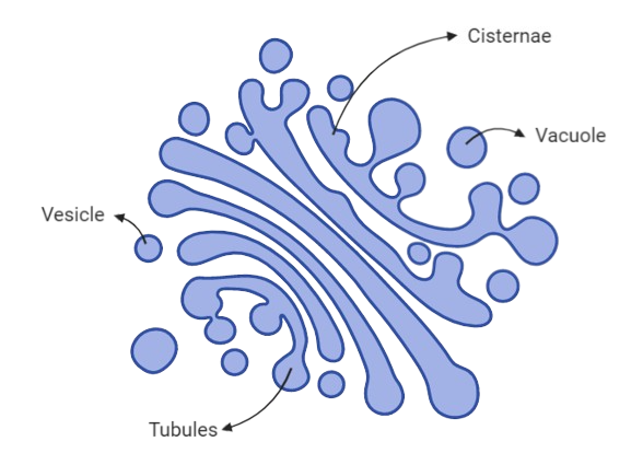
2.Tubules
- A complex array of associated vesicles and anastomosing tubules (30 to 50nm diameter) surround the dictyosome and radiate from it.
- In fact, the peripheral area of dictyosome is fenestrated (lace-like) in structure.
3.Vesicles
- The vesicles (60 nm in diameter) are of three types :
1.Transitional vesicles
- These are small membrane limited vesicles which are through to form as blebs from the transitional ER to migrate and converge to cis face of Golgi, where they coalesce to form new cisternae.
2.Secretory vesicles
- These are varied-sized membrane-limited vesicles which discharge from margins of cisternae of Golgi.
- They, often, occur between the maturing face of Golgi and the plasma membrane.
3.Clathrin-coated vesicles
- These are spherical protuberances, about 50mm in diameter and with a rough surface.
- They are found at the periphery of the organelle, usually at the ends of single tubules, and are morphologically quite distinct from the secretory vesicles.
- The clathrin-coated vesicles are known to play a role in intra-cellular traffic or membranes and of secretory products. i.e., between ER and Golgi, as well as between GELR region and the endosomal and lysosomal compartments.
Isolation and Chemical Composition
- Initially, Golgi apparatus was isolated only from cells of the epididymis, however in recent years, it has been isolated from number of plant and animal cells.
- The isolation of Golgi apparatus brough about mainly by gentle homogenization following by differential and gradient homogenization
- Gentle homogenization is preferred to preserve the stacks of cisternae,
- Due to its low density, Golgi apparatuses tend to form a distinct band in gradient centrifugation.
- The isolated Golgi apparatus is apparatus tend to form a distinct band in gradient centrifugation.
- The isolated Golgi apparatus washed with distilled water for purifying it, though, its secretory components are lost.
- Chemically, Golgi apparatus of rat liver contains about 60 per cent lipid material.
- The Golgi apparatus of animal cells contains phospholipids in the form of phosphatidyl choline, whereas, that of plant cells contains phosphatidic acid and phosphatidyl glycerol.
- The Golgi apparatus also contains a variety of enzyme, some of which have been used as cytochemical markers.
Cytochemical Properties of Golgi Apparatus
- Different parts of Golgi apparatus have been histochemically identified by specific staining properties:
1.Osmium tetraxide (OsO4)
- selectively impregnates the outer face (cis-face) of the Golgi apparatus.
- This stain adheres well to lipids, especially phospholipids and unsaturated fats.
2.Phosphotungstic acid (H3PO4.12WO3.24H2O).
- selectively stain the maturing or trans face of Golgi stack.
- This stain is an anionic stain having special affinity for polysaccharides and proteins.
3.Glycosyl tranferease and thiamine pyrophosphatase
- can be localized cyto-chemically in the trans cisternae of Golgi apparatus.
- Transferase enzymes are found to be located in the membrane of Golgi, not in the lumen of cisternae.
4.Acid phosphatase enzyme
- Is cyto-chemically marked in the GERL region.
Origin
- Origin of Golgi apparatus involves the formation of new cisternae and there is great variation in shape, number and size of cisternae in each stack (dictyosome).
- The process of formation of new cisternae may be performed by any of the following methods ;
- Individual stacks of cisternae may arise from the pre-existing stacks by division or fragmentation.
- The alternative method of origin of Golgi is based on denovo formation.
- In fact various cytological and biochemical envidences have established that the membranes of the Golgi apparatus are originated from the membranes of the smooth ER which in turn have originated from the rough ER.
- The proximal Golgi saccules are formed by fusion of ER-derived vesicles, while distal saccules “give their all” to vesicle formation and disappear.
- Thus, Golgi saccules are constantly and rapidly renewed.
- The cells of dormant seeds of higher plants generally lack Golgi apparatuses but they do display zone of exclusion having aggregation of small transition vesicles.
- Photomicrographs of cells in early stages of germination suggest progressive development of Golgi bodies in these zones of exclusion ; and the development of Golgi apparatuses coincides with the disappearance of the aggregation of vesicles.
Function
- Golgi vesicles are often, referred to as the ”traffic police” of the cell
- They play a key role in sorting many of the cell’s proteins and membrane constituents, and in directing them to their proper destinations.
- To perform this function, the Golgi vesicles contain different sets of enzymes in different types of vesicles-cis, middle and trans cisternae- that react with and modify secretory proteins passing through the Golgi lumen or membrane proteins and glycoproteins in the Golgi membranes as they are on route to their final destinations.
- For example a Golgi enzyme may add a “signal” or “tag” such as a carbohydrate or phosphate residues to certain proteins to direct them to their proper sites in the cell.
- Or, a proteolytic Golgi enzyme may cut a secretory or membrane protein into two or more specific segments (E.g., molecular processing involved in the formation of pancreatic hormone insulin: preproinsulin® proinsulin® insulin).
- Recently, in the function of Golgi apparatus, sub compartmentalization with a division of labour has been proposed between the cis region (in which proteins of RER are sorted and some of them are returned back possibly by coated vesicles), and the trans region in which the most refined proteins are further separated for their delivery to the various cell compartments (e.g., plasma membrane, secretory granules and lysosomes).
- Thus, Golgi apparatus is a centre of reception, finishing, packaging, and dispatch for a variety of materials in animal and plant cells :
1.Golgi Functions in Plants
- In plants, Golgi apparatus is mainly involved in the secretion of mainly involved in the secretion of materials of primary and secondary cell walls (e.g., formation and export of glycoprotein, lipids, pectins and monomers for hemicellulose, cellulose, lignin, etc).
- During cytokinesis of mitosis or meiosis, the vesicles originating from the periphery of Golgi apparatus, coalesce in the phragmoplast area to form a semisolid layer, called cell plate.
- The unit membrane of Golgi vesicles fuses during cell plate formation and becomes part of plasma membrane of daughter cells .