In-Situ Hybridization – Concept and Techniques
In Situ Hybridization
- In situ hybridization is another method for determining the chromosomal location of a particular gene.
- This method requires a DNA copy of the gene or its RNA product, which is used to make a molecule (called a probe) that is complementary to the gene of interest.
- The probe is made radioactive or is attached to a special molecule that fluoresces under ultraviolet (UV) light and is added to chromosomes from specially treated cells that have been spread on a microscope slide.
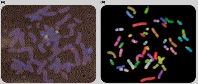
Figure In situ hybridization determine the chromosomal location of a gene. (a) FISH technique: in this case, the bound probe reveals sequences associated with the centromere. (b) SKY technique: 24 different probes, each specific for a different human chromosome and producing a different color, identify the different human chromosomes. (Courtesy of Dr. Hesed Padilla-Nash and Dr. Thomas Ried, NIH.)
- The probe binds to the complementary DNA sequence of the gene on the chromosome.
- The presence of radioactivity or fluorescence from the bound probe reveals the location of the gene on a particular chromosome (Figure).
- The use of fluorescence in situ hybridization (FISH) has been widely used to identify the chromosomal location of human genes.
- In Spectral Karyotyping (SKY) (Figure ), a set of 24 FISH probes, each specific to a different human chromosome and attached to a molecule that fluoresces a different color, allows each chromosome in a karyotype to be identified.
5.13
Figure 5.13 : In situ hybridization to locate specific genes on chromosomes. Here, six different DNA probes have been used to mark the location of their respective nucleotide sequences on human chromosome 5 at metaphase.
- The probes have been chemically labeled and detected with fluorescent antibodies. Both copies of chromosome 5 are shown, aligned side by side.
- Each probe produces two dots on each chromosome, since a metaphase chromosome has replicated its DNA and therefore contains two identical DNA helices. (Courtesy of David C. Ward.)
5.14
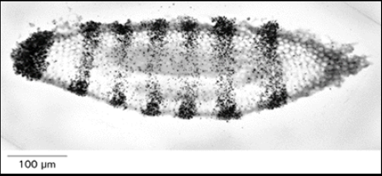
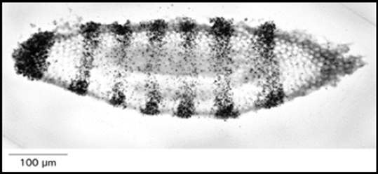
Figure 5.14 : In situ hybridization for RNA localization in tissues.
- Autoradiograph of a section of a very young Drosophila embryo that has been subjected to in situ hybridization using a radioactive DNA probe complementary to a gene involved in segment development.
- The probe has hybridized to RNA in the embryo, and the pattern of autoradiographic silver grains reveals that the RNA made by the gene (called ftz) is localized in alternating stripes across the embryo that are three or four cells wide.
- At this stage of development (cellular blastoderm), the embryo contains about 6000 cells. (From E. Hafen, A. Kuriowa, and W.J. Gehring, Cell 37:833-841, 1984. © Cell Press.)
In Situ Hybridization Techniques Locate Specific Nucleic Acid Sequences in Cells or on Chromosomes
- Nucleic acids, no less than other macromolecules, occupy precise positions in cells and tissues, and a great deal of potential information is lost when these molecules are extracted by homogenization.
- For this reason, techniques have been developed in which nucleic acid probes are used in much the same way as labeled antibodies to locate specific nucleic acid sequences in situ, a procedure called in situ hybridization.
- This can now be done both for DNA in chromosomes and for RNA in cells. Labeled nucleic acid probes can be hybridized to chromosomes that have been exposed briefly to a very high pH to disrupt their DNA base pairs.
- The chromosomal regions that bind the probe during the hybridization step are then visualized. Originally, this technique was developed using highly radioactive DNA probes, which were detected by autoradiography.
- The spatial resolution of the technique, however, can be greatly improved by labeling the DNA probes chemically instead of radioactively.
- For this purpose the probes are synthesized with special nucleotides that contain a modified side chain, and the hybridized probes are detected with an antibody (or other ligand) that specifically recognizes this side chain (Figure 5.13).
- In situ hybridization methods have also been developed that reveal the distribution of specific RNA molecules in cells in tissues.
- In this case the tissues are not exposed to a high pH, so the chromosomal DNA remains double-stranded and cannot bind the probe.
- Instead the tissue is gently fixed so that its RNA is retained in an exposed form that will hybridize when the tissue is incubated with a complementary DNA or RNA probe.
- In this way the patterns of differential gene expression can be observed in tissues.
- In the Drosophila embryo, for example, such patterns have provided new insights into the mechanisms that create distinctions between cells in different positions during development (Figure 5.14).