![]()
Flagellar Ultra structure : –
- Transmission electron microscope studies have shown that the bacterial flagellum is composed of three parts.
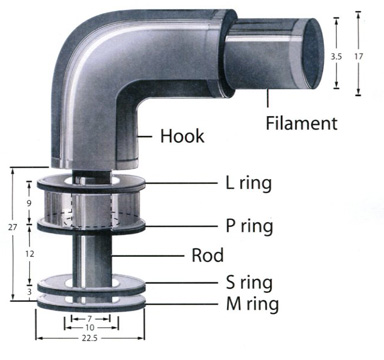
- (1) The longest and most obvious portion is the filament, which extends from the cell surface to the tip.
- (2) A basal body is embedded in the cell; and
- (3) a short, curved segment, the hook, links the filament to its basal body and acts as a flexible coupling.
- The filament is a hollow, rigid cylinder constructed of a single protein celled flagellin, which ranges in molecular weight from 30,000 to 60,000.
- The filament ends with a capping protein.
- Some bacteria have sheaths surrounding their flagella.
- For example Vibrio cholerae has a lipopolysaccharide sheath.
- The hook and basal body are quite different from the filament of different protein subunits.
- The basal body is the most complex part of a flagellum.
- In E.coli and most gram-negative bacteria, the body has four rings connected to a central rod.
- The outer L and P rings associate with the lipopolysaccharide and peptidoglycan layers, respectively.
- The inner M ring contacts the plasma membrane.
- Gram-positive bacteria have only two basal body rings, an inner ring connected to the plasma membrane and an outer one probably attached to the peptidoglycan.
Flagellar Synthesis
- The synthesis of flagella is a complex process involving at least 20 to 30 genes.
- Besides the gene for flagellin, 10 or more genes code for hook and basal body proteins ;
- other genes are concerned with the control of flagellar construction or function. It is not known how the cell regulates or determines the exact location of flagella.
The Mechanism of Flagellar Movement
- Procaryotic flagella operate differently from eukaryotic flagella.
- The filament is in the shape of a rigid helix, and the bacterium moves when this helix rotates.
- Considerable evidence shows that flagella act just like propellers on a boat.
- Bacterial mutants with straight flagella or abnormally long hook regions (polyhook mutants) cannot swim.
- When bacteria are tethered to a glass slide using antibodies to filament or hook proteins, the cell body rotates rapidly about the stationary flagellum.
- If polystyrene-latex beads are attached to flagella, the beads spin about the flagellar axis due to flagellar rotation.
- The flagellar motor can rotate very rapidly.
- The E. coli motor rotates 270 revolutions per second ; Vibrio alginolyticus averages 1,100 rps.
- The direction of flagellar rotation determines the nature of bacterial movement.
- Monotrichous, polar flagella rotate slowly clockwise.
- The rotating helical flagellar filament thrusts the cell forward in a run with the flagellum trailing behind.
- Monotrichous bacteria stop and tumble randomly by reversing the direction of flagellar rotation.
- Peritrichously flagellated bacteria operate in a somewhat similar way to move forward, the flagella rotate counterclockwise.
- As they do so, they bend at their hooks to form a rotating bundle that propels them forward.
- Clockwise rotation of the flagella disrupts the bundle and the cell tumbles.
- Because bacteria swim though rotation of their rigid flagella, there must be some sort of motor at the base.
- A rod or shaft extends from the hook and ends in the M ring, which can rotate freely in the plasma membrane.
- It is believed that the S ring is attached to the cell wall in gram positive cells and does not rotate .
- The P and L rings of gram negative bacteria would act as bearings for the rotating rod.
- There is some evidence that the basal body is a passive structure and rotates within a membrane embedded protein.
- The relationship of flagellar rotation to bacterial movement.
- The rotor is like an electrical motor turns in the center of a ring of electromagnets (the stator).
- The exact mechanism that drives basal drives basal body rotation still is not clear provides a more detailed depiction of the basal body in gram negative bacteria.
- The rotor portion of the motor seems to be made primarily of a rod, the M ring, and C ring joined to it on the cytoplasmic side of the basal body.
- These two rings are made of several proteins.
- The two most important proteins in the stator part of the motor are Mot A and Mot B.
- These form a proton channel thought the plasma membrane, and Mot B also anchors the Mot complex to cell wall peptidoglycan.
- There is some evidence that Mot A and G directly interact during flagellar rotation.
- This rotation is driven by proton or sodium gradients in prokaryotes, not directly by ATP as is the case with eukaryotic flagella.
- Bacteria can move by mechanisms other than falgellar rotations.
- Spriochetes are helical bacteria that travel through viscous substances such as mucus or mud by flexing and spinning movements caused by a special axial filament composed of periplasmic flagella.
- A very different type of motility, gliding motility, is employed by many bacteria : cyanobacteria .