![]()
Cytokinesis
- Mitosis is the process of separating the duplicates of each of the cell’s chromosomes.
- It is usually followed by division of the cell.
- However, there are cases (cleavage in the insect embryo is an example) where the chromosomes undergo the mitotic process without division of the cell.
- Thus a special term, cytokinesis, for the separation of a cell into two.
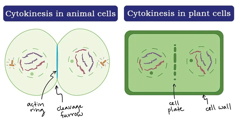
- In animal cells, a belt of actin filaments forms around the perimeter of the cell, midway between the poles.
- The interaction of actin and a myosin (not the one found in skeletal muscle) tightens the belt, and the cell is pinched into two daughter cells.
- In plant cells, a cell plate forms where the metaphase plate had been.
- The cell plate is synthesized by the fusion of multiple membrane-bounded vesicles.
- Their fusion supplies new plasma membrane for each of the two daughter cells.
- Synthesis of a new cell wall between the daughter cells then occurs at the cell plate.
- Cytokinesis is the process whereby the cytoplasm of a single eukaryotic cell is divided to form two daughter cells.
- It usually initiates during the late stages of mitosis, and sometimes meiosis, splitting a binucleate cell in two, to ensure that chromosome number is maintained from one generation to the next.
- In plant cells, a dividing structure known as the cell plate forms across the centre of the cytoplasm and a new cell wall forms between the two daughter cells.
Cell plate formation
- Due to the presence of a cell wall, cytokinesis in plant cells is significantly different from that in animal cells.
- Rather than forming a contractile ring, plant cells construct a cell plate in the middle of the cell.
- The Golgi apparatus releases vesicles containing cell wall materials.
- These vesicles fuse at the equatorial plane and form a cell plate.
- The cell plate begins as a fusion tube network, which then becomes a tubule-vesicular network (TVN) as more components are added.
- The TVN develops into a tubular network, which then becomes a fenestrated sheet which adheres to the existing plasma membrane.
- Phragmoplast and cell plate formation in a plant cell during cytokinesis. Left side:
- Phragmoplast forms and cell plate starts to assemble in the center of the cell.
- Towards the right: Phragmoplast enlarges towards the outside of the cell, leaving behind mature cell plate in the center.
- The cell plate will transform into the new cell wall once cytokinesis is complete.
- Cytokinesis in terrestrial plants occurs by cell plate formation.
- This process entails the delivery of Golgi-derived and endosomal vesicles carrying cell wall and cell membrane components to the plane of cell division and the subsequent fusion of these vesicles within this plane.
- After formation of an early tubule-vesicular network at the center of the cell, the initially labile cell plate consolidates into a tubular network and eventually a fenestrated sheet.
- The cell plate grows outward from the center of the cell to the parental plasma membrane with which it will fuse, thus completing cell division.
- Formation and growth of the cell plate is dependent upon the phragmoplast, which is required for proper targeting of Golgi-derived vesicles to the cell plate.
- As the cell plate matures in the central part of the cell, the phragmoplast disassembles in this region and new elements are added on its outside.
- This process leads to a steady expansion of the phragmoplast, and concomitantly, to a continuous retargeting of Golgi-derived vesicles to the growing edge of the cell plate.
- Once the cell plate reaches and fuses with the plasma membrane the phragmoplast disappears.
- This event not only marks the separation of the two daughter cells, but also initiates a range of biochemical modifications that transform the flexible cell plate into a cellulose-rich, stiff primary cell wall.
- The heavy dependence of cell plate formation on active Golgi stacks explains why plant cells, unlike mammalian cells, do not disassemble their secretion machinery during cell division.