![]()
Karyotype Analysis
- A group of plants or animals comprising a species is characterized by a set of chromosomes, have certain constant features.
- These features include chromosome number, size and shape of individual chromosomes and other attributes listed above.
- The term karyotype is given to the group of characteristics that identifies a particular chromosome set and is usually represented by a diagram called ideogram where chromosomes of haploid set of an organism are ordered in a series of decreasing size.
- The karyotypes of different groups are sometimes compared and similarities in karyotype are presumed to represent evolutionary relationships.
- Karyotype also suggest primitive or advanced feature of an organism.
This is Two type
(A) Symmetrical karyotype
- In this karyotyping all chromosome contain median and sub median centromere and the difference in size of chromosome is not very distinct.
- Plant having this type of karyotyping regarded as primitive
(B) Asymmetrical karyotyping –
- In this type of karyotyping the chromosome possesses subterminal centromere.
- These chromosomes depict a great variety in size i.e. from small to long chromosome are present in this asymmetrical karyotype.
- Plant having this karyotyping are regarded as advanced hence it can be concluded that karyotype of an individual or species is also a means for understanding the evolutionary process
- A symmetric and an asymmetric karyotype are shown in figure
- In 1931 G. A. Levitzky, a Russian scientist suggested that in flowering plants there is a predominant trend towards karyotype asymmetry.
- This trend has been carefully studied in the genus Crepis of the family compositae.
- In several cases it was shown that increased karyotype asymmetry was associated with specialized zygomorphic flowers.
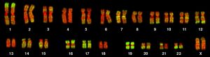
The karyotype of the human female contains 23 pairs of homologous chromosomes:
- 22 pairs of autosomes
- 1 pair of X chromosomes
The karyotype of the human male contains:
- the same 22 pairs of autosomes
- one X chromosome
- one Y chromosome
Process of Karyotyping
- A karyotype is a technique that allows geneticists to visualize chromosomes under a microscope.
- The chromosomes can be seen using proper extraction and staining techniques when the chromosomes are in the metaphase portion of the cell cycle.
- Detecting abnormalities is important for prenatal diagnosis, detection of carrier status for certain genetic diseases or traits, and for general diagnostic purposes.
Sterilization–
- It is an important step.
- All instruments and equipment’s should be properly sterilized.
Sampling and Culture–
- Karyotype analysis can be performed on virtually any population of rapidly dividing cells either grown in tissue culture or extracted from tumors.
- Chromosomes derived from peripheral blood lymphocytes are ideal because they can be analyzed three days after they are cultured.
- Lymphocytes can be induced to proliferate using a mitogen (a drug that induces mitosis) like phytohemagglutinin.
- Skin fibroblasts, bone marrow cells, chorionic villus cells,tumorcells, or amniocytes also can be used but require up to two weeks to obtain a sufficient amount of cells for analysis.
Cell Synchronization-
- The cultured cells are treated with colcemid, a drug that disrupts the mitotic spindle apparatus to prevent the completion of mitosis and arrests the cells in metaphase.
Harvesting and Slide preparation–
- The harvested cells are treated briefly with a hypotonic solution.
- This causes the nuclei to swell making it easier for technicians to identify each chromosome.
- The cells are fixed, dropped on a microscope slide, dried, and stained.
Observation in microscope –
- The most common stain used is the Giemsa stain.
- Other dyes, such as fluorescent dyes, can also be used to produce banding patterns.
Photography-
- Chromosome spreads can be photographed.
- Enlargement of photo and rearrangement to form Karyotype-
- The photographs are enlarged, cut out, and assigned into the appropriate chromosome number or they can be digitally imaged using a computer.
- In case of Human karyotype, there are seven groups (A-G) that autosomal chromosomes are divided into based on size and position of the centromere.
- The standard nomenclature for describing a karyotype is based on the International System.
Karyotype Evolution
- Two aspects of the process of speciation are of interest in the context of cytogenetics.
- The first of these is changes in ploidy i.e. changes in the number of the chromosomes, which themselves remain unaltered.
- Changes in ploidy can have both genetic and phenotypic effects, such as fertility changes, and can be used to great effecting plant breeding to produce new cultivars.
- Additional sets of chromosomes can be from the individual (autopolyploid) or from an organism of genetically distinct origin (allopolyploids).In the case of humans, changes in ploidy can have very severe consequences, as can the second process of interest: changes in karyotype.
- Karyotype changes can be thought of as being due to changes in either DNA content or chromosome structure as well as changes in chromosome numbers.
- In this context it is possible to see speciation and karyotype changes that are linked, as in the marsupials, or not linked, as in the hominids.
- Evolution and speciation are closely related to observable changes in an organism’s chromosomes.
- It should, however, be clearly borne in mind that karyotype changes are rarely enough for speciation to occur on their own. It is, after all, the phenotype expression of the genome which determines the position of the fine line, sometimes indefinable, between variation and speciation