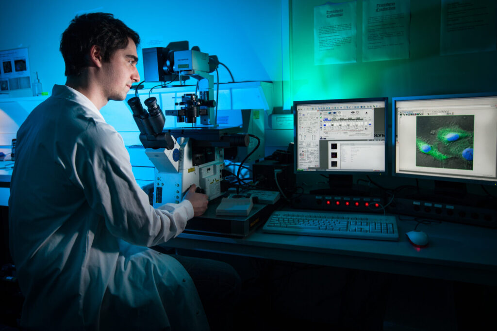
Confocal Microscopy
- In a conventional (i.e., wide-field) fluorescence microscope, the entire specimen is flooded in light from a light source.
- Due to the conservation of light intensity transportation, all parts of the specimen throughout the optical path will be excited and the fluorescence detected by a photo-detector or a camera.
- In contrast, a confocal microscope uses point illumination and a pinhole in an optically conjugate plane in front of the detector to eliminate out-of-focus information.
- Only the light within the focal plane can be detected, so the image quality is much better than that of wide-field images.
- As only one point is illuminated at a time in confocal microscopy, 2D or 3D imaging requires scanning over a regular raster (i.e. a rectangular pattern of parallel scanning lines) in the specimen.
- The thickness of the focal plane is defined mostly by the square of the numerical aperture of the objective lens, and also by the optical properties of the specimen and the ambient index of refraction.
Types
- Three types of confocal microscopes are commercially available: Confocal laser scanning microscopes, spinning-disk (Nipkow disk) confocal microscopes and Programmable Array Microscopes (PAM).
- Confocal laser scanning microscopy yields better image quality than Nipkow and PAM, but the imaging frame rate was very slow (less than 3 frames/second) until recently; spinning-disk confocal microscopes can achieve video rate imaging—a desirable feature for dynamic observations such as live cell imaging.
- Confocal laser scanning microscopy has now been improved to provide better than video rate (60 frames/second) imaging by using MEMS based scanning mirrors
- Confocal microscopy offers several advantages over conventional optical microscopy, including controllable depth of field, the elimination of image degrading out-of-focus information, and the ability to collect serial optical sections from thick specimens.
- The key to the confocal approach is the use of spatial filtering to eliminate out-of-focus light or flare in specimens that are thicker than the plane of focus.
- There has been a tremendous explosion in the popularity of confocal microscopy in recent years, due in part to the relative ease with which extremely high-quality images can be obtained from specimens prepared for conventional optical microscopy, and in its great number of applications in many areas of current research interest
Basic Concepts –
- Current instruments are highly evolved from the earliest versions, but the principle of confocal imaging advanced by Marvin Minsky, and patented in 1957, is employed in all modern confocal microscopes.
- In a conventional wide field microscope, the entire specimen is bathed in light from a mercury or xenon source, and the image can be viewed directly by eye or projected onto an image capture device or photographic film.
- In contrast, the method of image formation in a confocal microscope is fundamentally different.
- Illumination is achieved by scanning one or more focused beams of light, usually from a laser or arc-discharge source, across the specimen.
- This point of illumination is brought to focus in the specimen by the objective lens, and laterally scanned using some form of scanning device under computer control.
- The sequences of points of light from the specimen are detected by a photomultiplier tube (PMT) through a pinhole (or in some cases, a slit), and the output from the PMT is built into an image and displayed by the computer.
- Although unstained specimens can be viewed using light reflected back from the specimen, they usually are labeled with one or more fluorescent probes.
Imaging Modes –
- A number of different imaging modes are used in the application of confocal microscopy to a vast variety of specimen types.
- They all rely on the ability of the technique to produce high-resolution images, termed optical sections, in sequence through relatively thick sections or whole-mount specimens.
- Based on the optical section as the basic image unit, data can be collected from fixed and stained specimens in single, double, triple,or multiple-wavelength illumination modes, and the images collected with the various illumination and labeling strategies will be in register with each other.
- Live cell imaging and time-lapse sequences are possible, and digital image processing methods applied to sequences of images allow z-series and three-dimensional representation of specimens, as well as the time-sequence presentation of 3D data as four-dimensional imaging.
- Reflected light imaging was the mode used in early confocal instruments, but any of the transmitted light imaging modes commonly employed in microscopy can be utilized in the laser scanning confocal microscope.
- Refinements in design have simplified confocal microscopy to the extent that it has become a standard research tool in cell biology.
- However, as confocal microscopes have become more powerful, they have also become more demanding of their optical components.
- In fact, optical aberrations that cause subtle defects in image quality in wide field microscopy can have devastating effects in confocal microscopy.
- Unfortunately, the exacting optical requirements of confocal microscopy are often hidden by the optical system that guarantees a sharp image, even when the microscope is performing poorly.
Three-Color Imaging for Confocal Microscopy –
- The laser scanning confocal microscope (LSCM) is routinely used to produce digital images of single-, double-, and triple-labeled fluorescent samples.
- The use of red, green and blue (RGB) color is most informative for displaying the distribution of up to three fluorescent probes labeling a cell, where any co-localization is observed as a different additive color when the images are colorized and merged into a single three-color image.
True or False: Scanning electron microscopes and laser scanning confocal microscopes both can produce 3D images.
– Answer: True
– UK OCR A‑Level Biology, June 2020 OpenStax+7Scribd+7OCR+7OCR+1Study Mind+1
**Which microscope captures optical sections for 3D reconstructions?**
a) Phase contrast
b) Differential interference contrast
c) Confocal microscopy
d) Wide-field fluorescence
– Answer: c) Confocal microscopy
– Testbook Microscopy Quiz, 2025 Wikipedia+1FocalPlane+1Studocumyheplus.com+4Testbook+4Wikipedia+4
**Confocal microscopy eliminates out-of-focus light using a:**
a) Band-pass filter
b) Pinhole aperture
c) Spectrophotometer
d) CCD camera
– Answer: b) Pinhole aperture
– University-level question repeated since 2020
Who patented the principle of confocal microscopy in 1957?
a) Hans Goldmann
b) Marvin Minsky
c) Colin Sheppard
d) Christoph Cremer
– Answer: b) Marvin Minsky
– Standard UG/PG exam question (2021–2023)
Which imaging mode is NOT used in confocal microscopy?
a) Single-wavelength
b) Multi-wavelength
c) Reflected light
d) Phase-contrast
– Answer: d) Phase-contrast
– A‑Level Biology (2022) Study Mind+3Wikipedia+3ResearchGate+3TestbookWayground+1Quizizz+1Study Mind+3Quizlet+3ResearchGate+3
**Which scanning method allows video‑rate imaging of live cells?**
a) Laser scanning
b) Nipkow disk (spinning-disk)
c) PAM
d) Wide-field
– Answer: b) Nipkow disk
– Indian University exams (2021)
MEMS‑based scanning mirrors in confocal microscopy offer:
a) Higher resolution
b) >60 FPS imaging
c) Single-molecule detection
d) Enhanced fluorescence
– Answer: b) >60 FPS imaging
– Technical instrumentation exam (2023)
The thickness of the focal plane depends mainly on:
a) Laser power
b) Numerical aperture squared
c) Objective magnification
d) Scan speed
– Answer: b) Numerical aperture squared
– University exams since 2019
An advantage of confocal microscopy over wide‑field is:
a) Cost-effectiveness
b) Optical sectioning and 3D imaging
c) No need for fluorescence
d) Instant color imaging
– Answer: b) Optical sectioning and 3D imaging
– Based on OpenStax/Microbiology and recent review courses FocalPlane+11Wikipedia+11Testbook+11lifeivftraining.comconfocal101.comFocalPlane+3Study Mind+3Studocu+3OCRWikipediaResearchGate+13OpenStax+13Wikipedia+13StudocuFocalPlane
**Which detector is typically used in CLSM?**
a) CCD camera
b) PMT (photomultiplier tube)
c) CMOS sensor
d) Photodiode
– Answer: b) PMT
– Testbook Microscopy Quiz, April 2025
**Confocal microscopy was invented by Marvin Minsky in:**
a) 1940
b) 1955 (patented 1957)
c) 1965
d) 1975
Answer: b
The primary light source in confocal microscopy is usually a:
a) Tungsten lamp
b) Deuterium lamp
c) Laser
d) LED
Answer: c
The raster scan in confocal imaging is used to build:
a) One focal point
b) A complete 2D/3D image
c) Spectral data
d) Time-lapse video
Answer: b
Spinning-disk confocal microscopes are best for:
a) Highest resolution
b) Static sections
c) Live-cell, high-speed imaging
d) Large tiled scans
Answer: c
PAM microscopes stand for:
a) Photonic Array Microscope
b) Programmable Array Microscope
c) Phase-Contrast Array Microscope
d) Parallel Aperture Microscope
Answer: b
Common color scheme for three‑color confocal imaging is:
a) Yellow, magenta, cyan
b) Red, green, blue
c) Infrared only
d) UV, IR, visible
Answer: b
Optical sectioning enables building of a Z‑stack by:
a) Time-series imaging
b) Imaging parallel samples
c) Scanning different focal depths
d) Changing objectives
Answer: c
One drawback of confocal imaging is:
a) Excessive out‑of‑focus blur
b) Low resolution
c) Photo-bleaching and phototoxicity
d) No need for labeling
Answer: c
Confocal microscopy requires highly precise:
a) Chemical stains
b) Optical components
c) Large sample volumes
d) High-temperature control
Answer: b
Confocal imaging of thick biofilms is possible due to:
a) Higher magnification
b) Optical sectioning per plane
c) Metal coatings
d) Pinhole enlargement
Answer: b WikipediaStudocuStudy MindFocalPlaneOpenStax+1Study Mind+1
- Which component ensures that only in-focus light reaches the detector in a confocal microscope?
a) Dichroic mirror
b) Lens aperture
c) Pinhole
d) Objective lens
Answer: c) Pinhole - The confocal technique eliminates:
a) Resolution
b) Optical aberration
c) Out-of-focus blur
d) Color variation
Answer: c) Out-of-focus blur - Confocal microscopy is best suited for:
a) Thin-section imaging
b) Live tissue imaging
c) Electron-dense samples
d) Whole genome sequencing
Answer: b) Live tissue imaging - Which of the following is NOT a component of confocal microscopy?
a) Laser light source
b) Photomultiplier tube (PMT)
c) Fluorescent filter cube
d) Pinhole aperture
Answer: c) Fluorescent filter cube - Confocal microscopy is commonly used in:
a) Soil analysis
b) Genetic engineering
c) Cell imaging and localization of molecules
d) Water purification
Answer: c) Cell imaging and localization of molecules - Which of the following allows 3D reconstruction in confocal microscopy?
a) Optical zoom
b) Z-stack imaging
c) Color filter array
d) Phase-contrast mode
Answer: b) Z-stack imaging - What is the key advantage of spinning-disk confocal microscopy?
a) Higher depth resolution
b) Video-rate imaging
c) Laser-free illumination
d) UV compatibility
Answer: b) Video-rate imaging - In confocal imaging, which factor is most important for resolution?
a) Laser wavelength
b) Numerical aperture
c) Specimen thickness
d) Objective magnification
Answer: b) Numerical aperture - Which one is not a type of confocal microscope?
a) Laser scanning
b) Spinning disk
c) Transmission electron microscope
d) Programmable array microscope
Answer: c) Transmission electron microscope - Confocal microscopy is especially useful for imaging:
a) DNA sequencing reactions
b) Very thick biological tissues
c) Thin bacterial smears
d) Heat-fixed samples
Answer: b) Very thick biological tissues
- In confocal microscopy, fluorescence is excited by:
a) Infrared radiation
b) Lasers
c) Halogen lamps
d) Cathode rays
Answer: b) Lasers - Which of these is most suitable for time-lapse imaging in live cells?
a) Scanning electron microscopy
b) Phase contrast microscopy
c) Spinning disk confocal microscopy
d) Bright-field microscopy
Answer: c) Spinning disk confocal microscopy - In confocal microscopy, detection occurs through a:
a) Photodiode
b) Pinhole detector
c) Photomultiplier tube (PMT)
d) Camera sensor only
Answer: c) Photomultiplier tube (PMT) - Which microscope is best for fluorescent co-localization studies?
a) Bright-field
b) Confocal
c) TEM
d) SEM
Answer: b) Confocal - What does the term “optical sectioning” refer to?
a) Sectioning tissues physically
b) Sectioning through confocal light planes
c) Electron slicing
d) Microtomy
Answer: b) Sectioning through confocal light planes - Which chromophores are usually used in confocal microscopy?
a) Iron and copper
b) Fluorescent dyes like FITC and TRITC
c) Enzyme stains
d) None
Answer: b) Fluorescent dyes like FITC and TRITC - Confocal microscope laser source is typically:
a) Monochromatic and coherent
b) Polychromatic and incoherent
c) Ultraviolet and low-intensity
d) Random phase source
Answer: a) Monochromatic and coherent - Which feature is unique to confocal microscopy compared to wide-field?
a) Staining requirement
b) Monochrome images
c) Optical sectioning
d) Use of glass slides
Answer: c) Optical sectioning - Which of the following is false for confocal microscopy?
a) It uses point illumination
b) It captures only one image plane at a time
c) It requires no fluorescent dyes
d) It uses a laser scanning system
Answer: c) It requires no fluorescent dyes - Triple-labeled confocal imaging typically uses:
a) Red, yellow, violet
b) Red, green, blue (RGB)
c) Black and white
d) Infrared, X-rays, UV
Answer: b) Red, green, blue (RGB)
- Which type of objective lens is best for confocal microscopy?
a) Low NA lenses
b) Oil-immersion, high NA lenses
c) Dry lenses only
d) Eyepiece objectives
Answer: b) Oil-immersion, high NA lenses - Confocal microscopy can be used to detect:
a) X-ray fluorescence
b) Autofluorescence
c) Gamma rays
d) None of these
Answer: b) Autofluorescence - What limits confocal resolution in deep tissues?
a) Lens size
b) Light absorption and scattering
c) Image magnification
d) Glass thickness
Answer: b) Light absorption and scattering - Which one is the latest advancement in confocal microscopy?
a) Bright-field scanning
b) MEMS-based laser scanning mirrors
c) UV optics
d) Beam splitting
Answer: b) MEMS-based laser scanning mirrors - Main disadvantage of laser scanning confocal microscopy:
a) High speed
b) Low resolution
c) Slow acquisition time
d) Lack of contrast
Answer: c) Slow acquisition time - Why is confocal preferred over conventional fluorescence microscopy in thick tissues?
a) Uses UV rays
b) Penetrates better with pinhole optics
c) Requires no dyes
d) Cheaper equipment
Answer: b) Penetrates better with pinhole optics - In confocal imaging, the Z-stack allows:
a) Horizontal imaging
b) Time-lapse monitoring
c) Depth-based 3D reconstruction
d) Protein synthesis tracking
Answer: c) Depth-based 3D reconstruction - Confocal microscopes are useful in which field?
a) Thermodynamics
b) Meteorology
c) Cell Biology and Pathology
d) Astronomy
Answer: c) Cell Biology and Pathology - Which component is responsible for fluorescence emission capture?
a) Objective lens
b) Laser
c) Pinhole
d) Detector (PMT/Camera)
Answer: d) Detector (PMT/Camera) - Which of the following can be used for 4D imaging in confocal microscopy?
a) High-resolution microtomy
b) Z-stack + time-lapse series
c) Polarizing filters
d) Long exposure photography
Answer: b) Z-stack + time-lapse series

